CELENA® X – מערכת הדמיה אוטומטית ומדידה גבוהה (High Content Imaging)
מערכת ה‑CELENA® X High Content Imaging System של Logos Biosystems היא פתרון מתקדם המשלב דימות אוטומטי מלא וניתוח תוצאות מתוחכם במכשיר קומפקטי לשולחן המעבדה. מערכת זו מאפשרת עיבוד מהיר ואמין של תאי תרבית – מתא קפוא ועד דימות תאים חיים מורכב – כל זאת בעזרת ממשק אינטואיטיבי ואפשרות לבניית Workflows מותאמים אישית Logos Biosystems+1Advena Bio.
מאפיינים מרכזיים:
-
אוטומציה מלאה לזיהוי תאים ותנועה בין באריות וצלחות, עם שלב XYZ ממונע, מערכת פילטרים ניתנת להחלפה ומחליף עדשות אוטומטי Logos BiosystemsAdvena Bio.
-
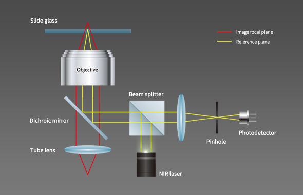
פוקוס מהיר ואמין, הודות לאופציות פוקוס אוטומטי מבוסס תמונה וכולל דוגמת לייזר, שמפחיתות את הפוטוטוקסיות ופוטובליצ’ינג Logos BiosystemsAdvena Bio.
-
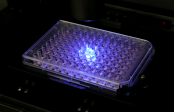
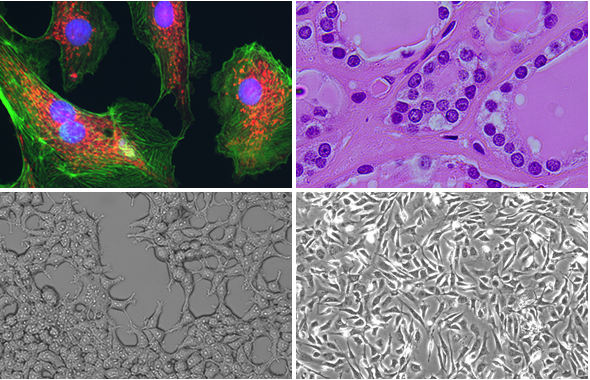
דימות ב‑4 ערוצים פלואורסצנטיים עם תמיכה ב‑Brightfield, Color Brightfield ו‑Phase Contrast, תאים קפואים וגם חיים, בצילומי Z‑stack ו‑Time‑Lapse Logos BiosystemsAdvena Bio.
-
תוכנת CELENA® X Cell Analyzer חזקה וגמישה, המאפשרת בניית פרוטוקולי ניתוח מותאמים אישית לבדיקות קבועות או ניסויים מתקדמים (כולל חי/מת, ספירה, מדידות צורתיות ועוד) Logos BiosystemsAdvena Bio.
-
תא אינקובציה On‑Stage, אידיאלי לניסויים בתנאים מבוקרים כמו טמפ’, לחות וריכוזי גזים שונים בזמן אמת Advena Bio.
ב־Lifegene, כיבואנית הרשמית של Logos Biosystems בישראל, תוכלו לקבל ייעוץ מותאם לצרכים שלכם, הדגמה מקצועית במעבדה, והדרכה מקיפה לשימוש במערכת CELENA® X. צרו עמנו קשר כדי לקחת את הניסוי שלכם לשלב הבא.
The CELENA® X High Content Imaging System is an integrated imaging system designed for rapid, high content image acquisition and analysis. Customizable imaging protocols, image-based and laser autofocusing modules, and a motorized XYZ stage simplify well plate imaging and slide scanning. The integrated CELENA® X Cell Analyzer software processes images and data for quantitative analysis. Analysis pipelines can be put together and reused to identify cellular or subcellular objects, process images for optimal data collection, and make various measurements. The CELENA® X is as flexible as it is powerful, with interchangeable objectives and filter cubes to accommodate a wide range of fixed and live cell imaging applications.
Fully Automated Plate And Slide Imaging
Automated vessel handling and scanning
Motorized XYZ stage, filter cube stage, and objective turret
Laser Autofocus
Rapid and reproducible focusing
Minimized phototoxicity and photobleaching
Live Cell Assay Support
Onstage incubation system for a variety of experiments in physiological and non-physiological conditions
Four Imaging Modes
Fluorescence imaging in four channels, brightfield, color brightfield, and phase contrast imaging
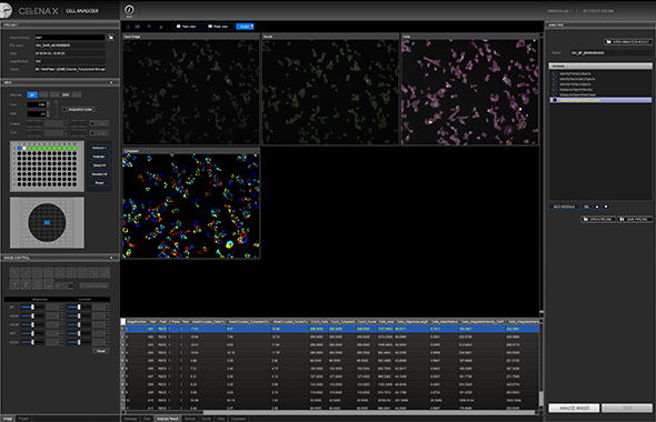
Powerful, Easy-To-Use User Interface
Simple setup of imaging protocols
Seamless integration of imaging and data analysis processes
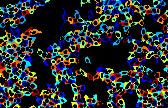
Customizable High Content Analysis
Create and customize image analysis projects
Quantitatively analyze multiple image-based phenotypes